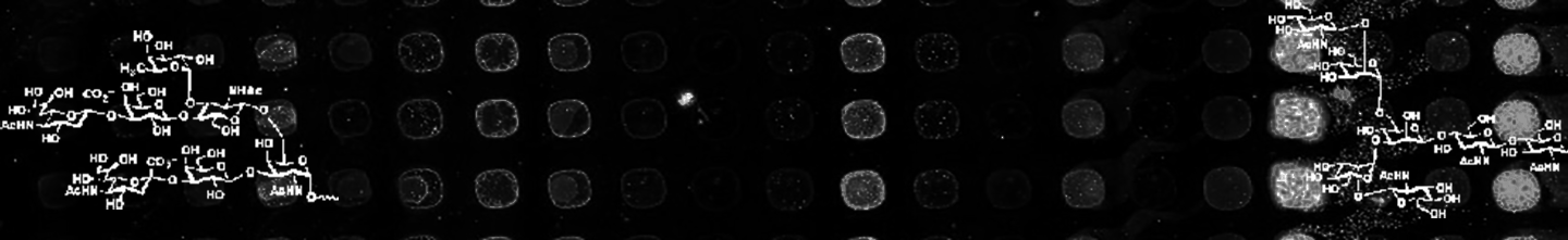
Lectin microarrays, a glycomic technology developed in the Mahal Lab, provide a rapid analysis of the glycome (1-3). These microarrays utilize immobilized carbohydrate-binding proteins at high spatial density to give specific information on the repertoire of glycans present, e.g. high mannose epitopes, branching patterns, and terminal α-2,3- or α-2,6-sialic acids, in a high-throughput format. By using lectin probes, we directly mimic how nature recognizes glycans, i.e. via 3-5 glycan binding epitopes, and we gain discrete structural information on glycan linkages. Unlike other glycomic methodologies such as mass spectrometry, lectin microarrays: 1) can directly observe the N-, O-linked and glycolipid glycomes simultaneously (2). 2) require minimal manipulation or derivitization of samples, allowing low amounts of raw starting material to be analyzed (100 μg or less of raw material is routinely used in our laboratory, compared to 3-5 mg for a typical glycomic mass spectrometry analysis) and 3) data is in the same format as mRNA and miRNA datasets, enabling facile data integration. Our dual-color methodology is unique to our laboratory and allows more accurate semi-quantitative analysis of the glycome (Fig.1, 2). We print our own lectin microarrays and include recombinant lectins from our laboratory (4,5). Our current print has over 100 lectins and antibodies. The discrete specificities of our probes are well characterized and many of them have been analyzed using the Consortium for Functional Glycomics glycan microarray (6). We have used our lectin microarray technology to identify prognostic glycosylation patterns in human esophageal cancer, validate GALNT7 as a pro-metastatic miRNA target, and study the role of glycosylation in HIV and microvesicle biogenesis, among other systems. This technology platform has enabled facile integration of glycomic information into multi’omic systems biology studies that are ongoing within the laboratory.
Lectin Microarray Analysis of Cervical Vaginal Lavage Fluid from (7)

References
- Pilobello, K.T.; Krishnamoorthy, L.; Slawek, D.; Mahal, L.K. Development of a Lectin Microarray for the Rapid Analysis of Protein Glycopatterns. ChemBioChem 2005, 6, 985-989.
- Pilobello, K.T.; Slawek, D.; Mahal, L.K. A ratiometric lectin microarray approach to analysis of the dynamic mammalian glycome, Proc. Natl. Acad. Sci., USA, 2007, 104, 10534-10539.
- Pilobello, K.T; Agrawal, P.; Rouse, R.; Mahal, L.K. Advances in lectin microarray technology: Optimized protocols for piezoelectric print conditions. Curr. Prot. Chem. Biol. 2013, 5, 1-23.
- Hsu, K.-L.; Gildersleeve, J.C.; Mahal, L.K. A simple strategy for the creation of a recombinant lectin microarray, Mol. BioSystems, 2008, 4, 654-662.
- Ribeiro, J.P.; Pau, W.K.; Pifferi, C.; Renaudet, O; Varrot, A; Mahal, L.K.‡; Imberty, A. ‡ Characterization of a high-affinity sialic acid specific CBM40 from Clostridium perfringens and engineering of a divalent form. Biochem. J. 2016, 473, 2109-18. doi: 10.1042/BCJ20160340. ‡ Co-corresponding authors.
- Wang, L.; Cummings, R.D.; Smith, D.F.; Huflejt, M.; Campbell, C.T.; Gildersleeve, J.D.; Gerlach, J.Q.; Kilcoyne, M.; Joshi, L.; Serna, S.; Reichardt, N.-C.; Pera, N.P.; Pieters, R.; Eng, W.S.; Mahal, L.K. Cross-Platform Comparison of Glycan Microarray Formats. Glycobiology, 2014, 24, 507-517. Doi: 10.1093/glycob/cwu019.
- Wang, L.; Koppolu, S.; Chappell, C.; Moncla, B.J.; Hillier, S.L.; Mahal, L.K. Studying the effects of reproductive hormones and bacterial vaginosis on the glycome of lavage samples from the cervicovaginal cavity. PLoS One, 2015, 10, e0127021. doi: 10.1371/journal.pone.0127021.